Microscope-Assisted Dentistry in Greater Kailash 1 | Tooth & Tonic
High-Magnification Precision Care, Micro-Endodontics & Conservative Tooth Preservation in GK 1
In conventional dentistry, diagnosis and clinical procedures are performed using the naked eye or low-power magnifying loupes (typically 2.5x to 4.5x magnification). While loupes offer an expanded view, human dental anatomy is microscopic: dentinal tubules measure merely micrometers across, accessory root canal pathways branch invisibly beneath the pulp floor, and structural micro-cracks (craze lines) often remain completely undetectable under ambient operatory lighting. When a clinician cannot clearly visualize microscopic anatomical variations, clinical compromises can occur—such as the unnecessary over-drilling of sound dentin, overlooking hidden bacterial canals, or failing to identify subgingival restorative margins.
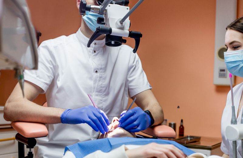
At Tooth & Tonic, located in Block R, Greater Kailash-1, restorative and endodontic care is guided by high-precision dental operating microscopes (DOM). By providing continuous, shadow-free coaxial LED illumination and stepless optical magnification ranging from 3x up to 25x, the operating microscope transforms clinical dentistry from tactile guesswork into direct, illuminated visualization. Our clinic applies microscopic principles across diagnostics, root canal therapy, cosmetic ceramic preparations, and microsurgery, ensuring that every intervention preserves maximum sound tooth structure and achieves sub-millimeter marginal accuracy.
Core Clinical Applications of Microscope-Assisted Dentistry at Our GK 1 Practice
Operating microscopes enhance visual acuity across every stage of dental diagnosis and intervention:
- Micro-Endodontics (Root Canal Therapy & Retreatment): Root canal systems feature intricate, branching channels. Under high magnification, our endodontists routinely identify hidden accessory canals (such as the mesiobuccal-2 or MB2 canal in upper molars), navigate calcified pulp chambers, remove separated instruments, seal root perforations with bioactive bioceramics, and preserve healthy pericervical dentin.
- Micro-Restorative & Biomimetic Cavity Preparation: Traditional drilling often removes excessive healthy tooth structure to ensure clear margins. Under magnification, our clinicians clear caries selectively with micro-burs, preserving healthy enamel prisms and establishing clean finish lines for composite bonding.
- Sub-Millimeter Prosthodontic Marginal Finishing: Fabricating monolithic zirconia crowns, ceramic onlays, and porcelain veneers requires precise finish lines (chamfers or shoulder margins). High-power magnification allows our dentists to smooth microscopic tooth borders, eliminating rough ledges and ensuring an airtight seal that prevents recurrent micro-leakage and gum inflammation.
- Early Detection of Micro-Fractures & Craze Lines: Structural micro-cracks in teeth can cause intermittent chewing discomfort that does not show up on standard 2D radiographs. The focused optical light of the microscope illuminates these fracture lines, allowing early preventive stabilization before a tooth fractures completely.
- Periodontal & Soft-Tissue Microsurgery: Using micro-instruments and fine ophthalmic sutures (6-0 to 8-0) under magnification, soft-tissue grafting and papilla-sparing surgical procedures heal with reduced trauma, minimal tissue disruption, and rapid post-operative vascularization.
The Optical Technology Behind the Dental Operating Microscope
A dental operating microscope represents an advanced optical system engineered specifically for the physical demands of oral microsurgery:
- Coaxial Shadowless Illumination: Unlike standard overhead operatory lights that cast shadows inside the oral cavity, the microscope projects focused, high-intensity LED light along the exact optical path of vision. This illuminates deep root canal chambers, narrow periodontal pockets, and posterior molar spaces.
- Apochromatic Optical Lenses: Medical-grade apochromatic glass lenses correct chromatic aberration (color fringing) and spherical distortion, delivering high color accuracy, contrast, and three-dimensional depth perception across all magnification tiers.
- Integrated High-Definition Video Documentation: Our operating microscopes are integrated with 4K digital video recording systems. This allows our clinicians to record high-resolution video and intraoperative photographs, providing patients with visual clarity regarding their dental conditions and procedural steps.
- Ergonomic Precision: Operating with a microscope requires upright posture, eliminating physical fatigue and allowing the clinician to execute fine micro-movements with steady motor control.
Schedule a Microscope-Assisted Dental Consultation in Greater Kailash 1
Experience high-magnification dental diagnosis, conservative restorations, and specialized micro-endodontics at Tooth & Tonic.
- Direct Call: 098216 54499
- WhatsApp Direct: Message Us on WhatsApp
- Clinic Address: R 98, Lower Ground Floor (LGF), Block R, Greater Kailash-1, New Delhi, Delhi 110048
- Location Details: Centrally located in Block R, Greater Kailash 1, minutes away from M-Block and N-Block markets, with reserved patient parking assistance and direct ground-level entry.
Step-by-Step Clinical Workflow with the Dental Microscope
- Diagnostic Positioning & Sterile Field Isolation: The patient is seated comfortably, and a dental rubber dam is placed over the target tooth. Rubber dam isolation provides a clean, dry field and prevents moisture from interfering with optical visualization.
- Parfocal Optical Calibration: The microscope is positioned, and the clinician calibrates the interpupillary distance, optical diopters, and fine focus to match the working distance.
- Low-Power Macro-Survey (3x–8x Magnification): We begin at lower magnification levels to assess the overall tooth architecture, soft-tissue boundaries, and broader occlusal landmarks.
- Mid-Power Precision Refinement (8x–16x Magnification): Used for conservative caries excavation, selective cavity conditioning, smoothing crown margins, and identifying primary root canal entrances.
- High-Power Micro-Navigation (16x–25x Magnification): Applied during delicate phases—such as scouting microscopic lateral canal openings, clearing calcifications, assessing apexes, or evaluating adhesive bonding interfaces.
- Digital Verification & Documentation: High-resolution intraoperative images are captured to verify the clean margins, complete canal debridement, or restorative seals.
Microscope-Assisted Dentistry vs. Conventional Dental Care
Clinical Parameter | Microscope-Assisted Dentistry (Tooth & Tonic GK 1) | Conventional Dentistry (Naked Eye / Basic Loupes) |
Magnification Range | Up to 20x – 25x stepless optical magnification | 1x (naked eye) to 2.5x – 4.5x (standard loupes) |
Illumination System | Direct coaxial, shadow-free LED light path | Indirect overhead chair light; frequent shadowing |
Dentin Conservation | Maximum preservation; selective micro-preparation | Higher risk of over-drilling sound tooth structure |
Canal Localization | Reliably locates accessory, lateral, and MB2 canals | Can miss hidden or calcified root canals |
Crack Detection | Detects hairline craze lines and micro-fractures | Micro-fractures often missed until tooth fractures |
Marginal Accuracy | Sub-millimeter fit; minimal gap margins | Larger tolerances; higher risk of open margins |
Long-Term Benefits of Microscopic Dental Care
Incorporating surgical magnification delivers tangible clinical advantages for patients:
- Long-Term Tooth Survival: Conserving natural enamel and pericervical dentin keeps teeth mechanically resilient against functional chewing loads over time.
- Lower Retreatment Rates: Illuminating and sanitizing every complex canal pathway during root canal therapy minimizes residual bacterial colonies, reducing the likelihood of persistent infections or retreatment.
- Predictable Restorative Longevity: Restorations bonded to smooth, precise microscopic margins seal out oral fluids and bacteria, preventing secondary cavities and recurrent decay.
- Conservative Interventions: High magnification allows our team to address early micro-demineralization and tiny cracks before they require extensive crowns or surgical extractions.
Frequently Asked Questions for Greater Kailash 1 (GK 1) Patients
What is microscope-assisted dentistry?
Microscope-assisted dentistry involves utilizing advanced dental operating microscopes that magnify the oral cavity up to 25 times its natural size while providing direct, shadow-free illumination. This allows our clinicians to perform diagnostic, restorative, and root canal procedures with exceptional precision and conservation of natural tooth structure.
Why is an operating microscope necessary for root canal treatment?
Root canal channels are narrow, curved, and often branch into microscopic accessory pathways (such as the MB2 canal in molars) that cannot be seen with the naked eye. The microscope illuminates these pathways, allowing the endodontist to thoroughly clean, shape, and seal the entire canal system while preserving the tooth’s structural dentin.
Is a dental operating microscope used for all dental procedures at your GK 1 practice?
Our clinicians utilize the microscope for procedures where high magnification provides clear clinical benefits—including complex root canals, endodontic retreatments, sub-millimeter crown preparation, delicate veneer finishing, micro-crack diagnostics, and soft-tissue microsurgery.
Does microscope-assisted dentistry make the appointment uncomfortable?
No. The microscope is positioned several inches above your face and does not touch you. All clinical care is performed under targeted localized numbing to ensure you remain completely relaxed and comfortable throughout the visit. Because procedures are minimally invasive, post-operative recovery is often quicker.
Can the microscope detect cracked teeth that do not appear on X-rays?
Yes. Early hairline cracks (craze lines and cracked tooth syndrome) often run vertically or horizontally without enough separation to appear on standard 2D dental X-rays. Under high magnification and focused coaxial illumination, these micro-cracks become visible, allowing for early treatment before the tooth fractures through the root.
Where is Tooth & Tonic located in Greater Kailash 1?
Tooth & Tonic is located at R 98, Lower Ground Floor (LGF), Block R, Greater Kailash-1, New Delhi 110048. Our clinic is centrally positioned within GK 1, just a short 3- to 5-minute drive from M-Block Market and N-Block Market. We routinely care for patients from nearby South Delhi areas including Greater Kailash 2 (GK2), Kailash Colony, East of Kailash, Defence Colony, Chittaranjan Park (CR Park), Pamposh Enclave, and Nehru Place. Reserved patient parking and valet assistance are available on arrival.
Does microscope-assisted dentistry take longer than conventional treatments?
Initial diagnostic inspection and preparation may take slightly more time due to the high level of detail involved. However, because procedures are performed with visual precision on the first attempt, the need for repeated appointments, corrective adjustments, and subsequent retreatments is significantly reduced.