Digital RVG X-Ray Diagnostics in Greater Kailash 1 | Tooth & Tonic
High-Resolution Digital Intraoral Radiography, Ultra-Low Radiation Imaging & Immediate Chairside Diagnostics in GK 1
Accurate dental treatment relies on clear diagnostic visualization. While visual examinations and optical magnification reveal surface enamel conditions, many dental problems develop beneath the surface—within the internal pulp chamber, between tight interdental contact areas, along the root canals, and inside the supporting alveolar bone. Conventional dental radiography previously required traditional photographic film, long radiation exposure times, and chemical developing solutions that took minutes to process and generated chemical waste.
At Tooth & Tonic, located in Block R, Greater Kailash-1, intraoral dental imaging is powered by advanced RVG (Radio-Visio-Graphy) digital sensors. RVG technology replaces traditional dental film with high-sensitivity electronic sensors. When exposed to a focused micro-beam of X-rays, the sensor captures the image and transmits it to chairside monitors in seconds. This digital approach reduces radiation exposure by up to 80% to 90% compared to traditional film, providing crisp, high-contrast imagery that aids in identifying early dental decay, evaluating bone support, and verifying root canal procedures.
Advanced RVG Sensor Technology at Our GK 1 Practice
Our Greater Kailash 1 facility utilizes modern digital intraoral sensors engineered for diagnostic clarity, patient comfort, and radiation safety:
- CMOS & Scintillator Technology: Our digital RVG sensors use advanced Complementary Metal-Oxide-Semiconductor (CMOS) chips paired with optical scintillators. This configuration converts X-ray photons into clear digital signals with high line-pair resolution, capturing fine details such as micro-fractures, accessory root canals, and early enamel demineralization.
- Up to 90% Radiation Reduction (ALARA Standard): Because digital sensors are far more sensitive to X-ray photons than traditional silver halide film, the required radiation exposure time is reduced to milliseconds. We strictly follow the ALARA (As Low As Reasonably Achievable) safety principle, making digital X-rays safe for children, adults, and seniors.
- Ergonomic Rounded Sensor Geometry: Traditional film packets had sharp, rigid edges that could press uncomfortably against the floor of the mouth or palate. Our modern digital sensors feature smooth, rounded corners and thin profiles, minimizing gag reflexes and soft-tissue irritation.
- Real-Time Chairside Image Processing: Images appear on our operatory monitors within seconds of capture. Clinicians can magnify specific root areas, adjust contrast and brightness, invert grayscale tones, and take calibrated digital measurements without repeating exposures.
- Eco-Friendly & Fully Digital: Digital radiography eliminates the hazardous chemical developing agents (fixers and developers) and lead foil waste associated with traditional darkrooms, integrating seamlessly into our green dental workflow.
Key Clinical Applications of Digital RVG X-Rays
RVG intraoral radiographs serve as a routine diagnostic tool across our practice disciplines in Greater Kailash 1:
- Detecting Interproximal Dental Caries: Cavities forming between tight back molars are often hidden during visual checks. Bitewing RVG radiographs reveal enamel breakdown and dentin involvement early, allowing for conservative composite restorations before the pulp is compromised.
- Root Canal Treatment (Endodontics): RVG imaging is essential at each step of a root canal. It helps our endodontists confirm diagnostic working length, verify file positioning inside root tips, evaluate disinfection boundaries, and verify the dense three-dimensional seal (obturation) of the canals.
- Periodontal Bone Level Evaluation: Periapical RVG radiographs illustrate the crestal alveolar bone height around individual teeth, helping clinicians identify vertical or horizontal bone loss caused by gum disease.
- Evaluating Restorative Margins: Checking the precision fit of crowns, bridges, inlays, and composite fillings. RVG images detect microscopic marginal overhangs or gaps where bacterial plaque could trigger secondary decay.
- Assessing Dental Trauma: In cases of chipped or knocked teeth from sports or accidents, RVG radiographs assess root integrity, check for root fractures, and evaluate the periodontal ligament space.
- Pediatric Dental Development: Evaluating milk tooth root resorption, assessing developing permanent tooth buds beneath the gums, and diagnosing early childhood caries.
Schedule Your Digital Dental Check-Up in Greater Kailash 1
Benefit from fast, ultra-low-radiation digital X-ray diagnostics and comprehensive care at Tooth & Tonic.
- Direct Call: 098216 54499
- WhatsApp Inquiries: Connect via WhatsApp
- Clinic Address: R 98, Lower Ground Floor (LGF), Block R, Greater Kailash-1, New Delhi, Delhi 110048
- Location Details: Centrally located in Block R, Greater Kailash 1, minutes away from M-Block and N-Block markets, with reserved patient parking assistance and direct ground-level entry.
Step-by-Step RVG Imaging Protocol at Tooth & Tonic GK 1
- Protective Preparation: Before capturing an image, a protective lead apron equipped with a thyroid collar is positioned over your chest and neck to shield your body from secondary scatter radiation.
- Sensor Hygienic Barrier: A fresh, single-use medical-grade hygienic barrier sleeve is placed over the digital sensor to maintain strict infection control standards.
- Precise Sensor Positioning: The sensor is placed comfortably inside the mouth adjacent to the target tooth, stabilized with a color-coded positioning ring (Rinn holder). This parallel-technique holder ensures an accurate right-angle beam alignment, preventing elongation or foreshortening distortion.
- Instant Micro-Exposure: The handheld or wall-mounted digital X-ray unit is aligned with the positioning ring. A fraction-of-a-second exposure is triggered.
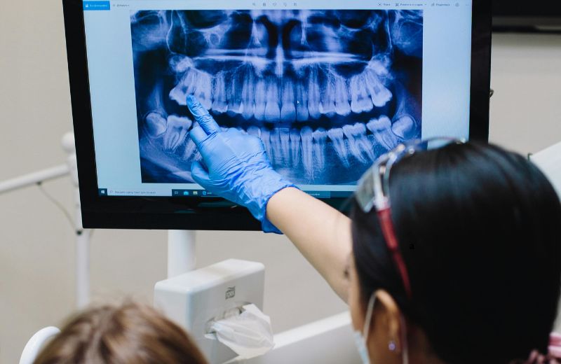
- Chairside Digital Analysis: The radiograph displays immediately on the monitor above your dental chair. Our clinician reviews the high-resolution image with you, explaining anatomical findings, pointing out areas of concern, and answering your questions clearly.
Diagnostic Comparison: RVG vs. OPG vs. 3D CBCT
Imaging Modality | Digital RVG X-Ray | Panoramic OPG X-Ray | In-House 3D CBCT Scan |
Field of View | Highly localized (2 to 3 teeth) | Full mouth (both jaws, sinuses, TMJ) | Targeted or full-jaw volumetric 3D |
Perspective | 2D high-resolution close-up | 2D broad continuous scan | 3D volumetric cross-sections |
Sensor Position | Intraoral (inside the mouth) | Extraoral (outside the mouth) | Extraoral (outside the mouth) |
Resolution Level | Exceptionally high detail resolution | Moderate overview resolution | High-definition cross-sectional detail |
Best Used For | Cavities, root canals, crown margins | Wisdom teeth, broad screening, TMJ | Implants, bone grafting, complex roots |
Safety and Radiation Facts
Patients frequently inquire about radiation exposure from dental X-rays. Because our clinic uses modern digital RVG sensors:
- A single digital RVG exposure delivers an ultra-low effective dose of approximately 0.001 to 0.005 mSv.
- This amount is comparable to the natural background radiation you encounter during a standard 1- to 2-hour commercial flight or a few hours of normal daily living outdoors.
- Targeted collimators restrict the micro-beam strictly to the dimensions of the digital sensor, preventing scatter radiation to surrounding tissues.
Frequently Asked Questions for Greater Kailash 1 (GK 1) Patients
How safe are digital RVG X-rays compared to old film X-rays?
Digital RVG sensors require significantly less radiation—typically 80% to 90% less than older film methods—because the digital sensor is much more sensitive to X-rays than physical film. Combined with protective lead aprons and thyroid shields, radiation exposure is kept to an absolute minimum.
Can pregnant patients receive an RVG X-ray at your GK1 clinic?
Preventative dental check-ups can often be scheduled around pregnancy, but urgent dental conditions (such as acute infections or toothaches) require prompt diagnosis. When medically indicated, digital RVG X-rays can be performed with double lead apron protection. Because the focused micro-beam is directed only at the mouth and the exposure lasts milliseconds, scatter radiation to the abdomen is negligible.
What if I have a strong gag reflex when the sensor is placed in my mouth?
Our digital sensors have rounded, ergonomic borders designed for comfort. We utilize specialized positioning holders that seat the sensor securely without slipping. If you have a sensitive gag reflex, our team will guide you through simple breathing techniques, apply a localized desensitizing spray to the palate, or use our extraoral panoramic OPG scanner as a comfortable alternative.
How long does an RVG X-ray take to display on screen?
Unlike traditional dental film that required 5 to 10 minutes of chemical processing in a darkroom, digital RVG radiographs appear on our chairside screens within 3 to 5 seconds.
Why does my dentist need an X-ray if my tooth looks fine on the outside?
Visual examinations only show the outer enamel surfaces. Many dental problems—such as decay between teeth, infections around root tips, internal resorption, cysts, and bone loss—develop beneath intact enamel. An RVG radiograph allows our team to identify and address issues early, before they compromise the tooth structure.
Where is Tooth & Tonic located in Greater Kailash 1?
Tooth & Tonic is located at R 98, Lower Ground Floor (LGF), Block R, Greater Kailash-1, New Delhi 110048. Our clinic is centrally positioned within GK 1, just a short 3- to 5-minute drive from M-Block Market and N-Block Market. We conveniently serve patients from nearby South Delhi localities including Greater Kailash 2 (GK2), Kailash Colony, East of Kailash, Defence Colony, Chittaranjan Park (CR Park), Pamposh Enclave, and Nehru Place. Reserved patient parking assistance is available on arrival.
Can digital RVG images be emailed to me for my personal records?
Yes. Because all RVG radiographs are stored electronically in high-resolution digital formats, we can securely email your diagnostic images to you or provide them for external specialist consultations whenever needed.