Digital OPG Dental X-Ray in Greater Kailash 1 | Tooth & Tonic
In-House Full-Mouth Panoramic Dental Radiography, Low-Dose Digital Imaging & Jaw Diagnostics in GK 1
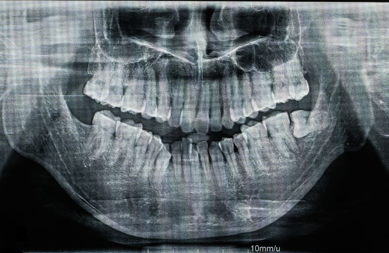
Comprehensive dental diagnosis often requires a broad, panoramic perspective of the entire maxillofacial region. While small intraoral periapical X-rays (RVG) provide high-resolution views of isolated individual teeth, they capture only two or three teeth at a time. An Orthopantomogram (OPG Dental X-Ray), commonly referred to as a panoramic dental X-ray, provides an expansive, continuous two-dimensional panoramic visualization of the entire oral framework in a single exposure.
An OPG scan captures the upper jaw (maxilla), lower jaw (mandible), all 32 erupted and unerupted teeth, temporomandibular joints (TMJ), maxillary sinuses, and the surrounding alveolar bone structures. It is an essential diagnostic screening tool for identifying hidden dental conditions that lie beneath the surface of the gums and within the deeper jawbone architecture.
At Tooth & Tonic, located in Block R, Greater Kailash-1, diagnostic radiography is performed entirely in-house using modern digital panoramic imaging systems. Patients visiting our GK 1 clinic do not have to travel across South Delhi to external diagnostic imaging labs or wait days for reports. We capture, process, and analyze high-definition panoramic images chairside in minutes, enabling prompt, coordinated treatment planning.
Advanced Panoramic Technology at Our GK 1 Practice
Our Greater Kailash 1 digital radiography suite utilizes high-definition digital sensor technology engineered for diagnostic accuracy and low radiation exposure:
- Digital Panoramic Sensors (CMOS/CCD): Moving away from traditional film-based radiography that required chemical developing agents, our digital OPG unit utilizes direct digital sensors. This produces high-contrast, high-resolution imagery instantly on chairside monitors, allowing for magnification, contrast adjustment, and measurement calibration.
- Focal Trough Auto-Correction: The focal trough is the curved three-dimensional image layer where anatomical structures appear sharpest. Our digital unit adjusts its path to match the unique curvature of your jaw, reducing edge blurring and ghost shadows.
- ALARA Low-Dose Radiation Protocol: By following the clinical principle of ALARA (As Low As Reasonably Achievable), our digital panoramic scanner delivers a fraction of the radiation exposure associated with traditional dental film radiography, ensuring a safe diagnostic environment for children, adults, and seniors.
- Completely Extraoral & Non-Invasive: For patients who have a severe, sensitive gag reflex, small oral openings, or acute mouth pain, an OPG is completely external. No sensors, film packets, or plastic holders are placed inside your mouth.
- Integrated Multi-Disciplinary Workflows: Digital OPG images are stored as universal digital files, allowing instant sharing with our in-house team of orthodontists, oral surgeons, implantologists, and endodontists.
Key Clinical Indications for an OPG Scan
An OPG X-ray is recommended across multiple clinical scenarios at our Greater Kailash 1 practice:
- Impacted Wisdom Teeth (Third Molars): Evaluating the position, angulation, and root depth of unerupted or partially erupted wisdom teeth, including their relationship to the inferior alveolar nerve canal.
- Orthodontic Treatment Planning: Assessing overall dental development, tooth root parallelity, crowding patterns, and missing or extra teeth (supernumeraries) before initiating clear aligners or fixed braces.
- Periodontal Bone Loss Screening: Measuring generalized bone support around all teeth to determine the extent and pattern of periodontal bone resorption caused by chronic gum disease.
- Screening for Cysts, Tumors & Jaw Lesions: Detecting asymptomatic odontogenic cysts, bone infections, calcifications, and bony abnormalities that can develop silently inside the jawbone.
- Temporomandibular Joint (TMJ) Evaluation: Examining the symmetry, shape, and bone health of the condylar heads in patients experiencing jaw clicking, stiffness, or bruxism.
- Dental Implant & Bone Assessment: Serving as an initial panoramic screening tool to evaluate available bone height before detailed 3D CBCT scans are performed.
- Dental Trauma & Fractures: Assessing mandibular or alveolar bone fractures following accidental falls, sports impacts, or facial trauma.
Schedule Your Digital OPG X-Ray in Greater Kailash 1
Benefit from rapid, in-house digital panoramic dental imaging and comprehensive diagnostic care at Tooth & Tonic.
- Direct Call: 098216 54499
- WhatsApp Inquiries: Connect via WhatsApp
- Clinic Address: R 98, Lower Ground Floor (LGF), Block R, Greater Kailash-1, New Delhi, Delhi 110048
- Convenient Access: Located in Block R, Greater Kailash 1, minutes away from M-Block and N-Block markets, with reserved patient parking assistance and direct ground-level entry.
What to Expect During an OPG X-Ray at Tooth & Tonic GK 1
The digital panoramic imaging process is swift, open-air, and entirely comfortable:
- Preparation & Metal Removal: You will be requested to remove any metallic objects above the neck—such as spectacles, hairpins, earrings, nose rings, tongue studs, neck chains, and removable partial dentures—to prevent metallic artifacts from obscuring diagnostic areas.
- Protective Shielding: A lightweight protective lead apron with a thyroid collar is provided to shield the rest of your body from secondary scatter radiation.
- Ergonomic Positioning: You will stand or sit upright comfortably inside our open, spacious panoramic unit. Unlike medical CT or MRI tunnels, the machine is completely open-air.
- Alignment & Bite-Peg Guide: You will gently rest your chin on a contoured chin rest and lightly bite onto a small, sterile, disposable plastic bite-peg to align your front teeth within the focal trough. Side temple supports stabilize your head to prevent minor movement.
- Scan Acquisition: The rotating arm of the OPG unit circles smoothly and quietly around your head for roughly 12 to 15 seconds. You simply remain still, keep your tongue pressed against the roof of your mouth, and breathe normally through your nose.
- Instant Digital Evaluation: The digital image appears immediately on our operatory monitors, allowing our dental team to review findings and discuss treatment steps with you on the spot.
Diagnostic Comparison: OPG vs. RVG vs. 3D CBCT
Diagnostic Metric | Panoramic OPG X-Ray | Intraoral Digital RVG X-Ray | 3D CBCT Scan |
Field of View | Full mouth (both jaws, all teeth, TMJ) | Localized (2 to 3 teeth per image) | Targeted or full-jaw volumetric 3D |
Dimension | 2-Dimensional panoramic view | 2-Dimensional flat projection | 3-Dimensional cross-sectional view |
Sensor Location | Extraoral (outside the mouth) | Intraoral (placed inside the mouth) | Extraoral (rotates around head) |
Patient Experience | Easy; zero gag reflex | May trigger gagging in back teeth | Open-air; zero gag reflex |
Primary Clinical Use | Baseline overview, wisdom teeth, TMJ | Root canals, small interproximal cavities | Implant surgery, complex impactions |
The Advantage of In-House Imaging in Greater Kailash 1
Many traditional dental practices lack integrated radiographic equipment, requiring patients to book separate appointments at external medical laboratories across South Delhi. This delays consultations, creates diagnostic disconnects, and adds unnecessary travel.
At Tooth & Tonic GK 1, having advanced OPG, 3D CBCT, and RVG technology within our facility ensures:
- Zero Wait Times: Diagnostic imaging is completed during your primary appointment.
- Immediate Clinical Correlation: Your dentist directly inspects the radiograph alongside your clinical examination.
- Collaborative Care: Multi-specialty opinions—from our oral surgeons to orthodontists—occur chairside in real time.
- Digital Records: High-resolution digital imaging files can be shared securely with you for your personal medical records.
Frequently Asked Questions for Greater Kailash 1 (GK 1) Patients
Do I have to visit an outside diagnostic laboratory for an OPG scan?
No. Tooth & Tonic features an advanced in-house digital OPG and 3D imaging suite directly inside our clinic at R 98, Block R, Greater Kailash 1. Your panoramic X-ray is captured and reviewed on-site during your visit.
Is an OPG X-ray safe in terms of radiation exposure?
Yes. Our clinic uses digital panoramic sensors that emit very low levels of radiation—significantly less than traditional film X-rays and a tiny fraction of a medical chest X-ray or medical CT scan. We strictly follow ALARA safety protocols, providing lead aprons for complete radiation protection.
Can an OPG detect impacted wisdom teeth and jaw cysts?
Yes. An OPG is one of the most effective screening tools for impacted third molars. It displays the entire mandible and maxilla in one image, revealing whether wisdom teeth are growing horizontally, impinging on adjacent teeth, or associated with dentigerous cysts or infections.
Is an OPG X-ray suitable for patients with a severe gag reflex?
Yes. Because the OPG is completely extraoral, no sensor or plastic device is inserted into your mouth. You simply rest your chin on an external support while the machine rotates around your head, making it ideal for patients with a sensitive gag reflex, children, and elderly individuals.
Can external dentists refer patients to Tooth & Tonic for an OPG?
Yes. We routinely provide digital radiographic services for referring dentists and oral surgeons across Greater Kailash and South Delhi. Digital panoramic images are provided promptly via email or digital storage.
Where is Tooth & Tonic located in Greater Kailash 1?
Tooth & Tonic is located at R 98, Lower Ground Floor (LGF), Block R, Greater Kailash-1, New Delhi 110048. Our clinic is centrally positioned within GK 1, just a 3- to 5-minute drive from M-Block Market and N-Block Market. We conveniently serve patients from nearby South Delhi localities including Greater Kailash 2 (GK2), Kailash Colony, East of Kailash, Chittaranjan Park (CR Park), Defence Colony, Pamposh Enclave, and Nehru Place. Reserved patient parking assistance is available on arrival.
How long does an OPG X-ray appointment take?
The scanning process itself takes only 12 to 15 seconds. The entire appointment—including positioning, protective apron placement, image acquisition, and chairside review with our dental team—typically takes 10 to 15 minutes.